The lighting community is currently considering the development and use of metrics that characterize circadian light—or light that acts as a stimulus for the human circadian system—to establish a design-performance standard (for a particular time of day) and to further research the suppression of melatonin production. Both light and dark set the timing of the master clock in our brains’ suprachiasmatic nuclei (SCN), and disruption of this clock has been shown to negatively affect many health outcomes, from breast cancer to diabetes.
Among the proposed metrics is “melanopic lux,” a term that can be loosely described as flux density weighted not by the photopic luminous efficiency function—or V(λ), which peaks at 555 nanometers and is based on the response of foveal, long- and middle-wavelength sensitive cones—but by a luminous efficiency function, which peaks at 480 nanometers and is based on the action spectrum of melanopsin. (Melanopsin is the retinal photopigment within our eyes’ intrinsically photosensitive retinal ganglion cells, or ipRGCs, which form the main neural conduit from the retina to the brain’s master clock.)
Lighting Research Center
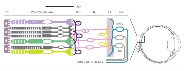
Detail of retina in the human eye
Photometric units have not been established for the circadian luminous efficiency function; consequently, quantifying light in terms of melanopic lux has yet to be defined. More importantly, the impact on the SCN by different levels of melanopic lux is completely unknown. It is thus impossible to use the action spectrum of melanopsin to describe the effectiveness of electric light or daylight for stimulating the human circadian system.
Photometric units have not been established for the luminous efficiency function; consequently, quantifying light in terms of melanopic lux has yet to be defined.
Metrics Matter
Apart from units and quantities, basing a metric on melanopsin alone is fundamentally problematic. With the exception of scotopic vision, a photopigment action spectrum is never the same as the spectral sensitivity of a neural channel response measured with electrophysiological or behavioral methods. After absorbing light, a photopigment generates a neural response from the photoreceptor. These responses are then combined into neural channel responses. For example, V(λ) reflects the combined photon absorptions of erythrolabe and chlorolabe, which generate long- and short-wavelength cone responses, respectively. (Erythrolabe is the pigment in our retinal cones that is most sensitive to the red range of the spectrum, while chlorolabe is the pigment most sensitive to the green range.)
Glossary
Suprachiasmatic nuclei (SCN)—site of the master clock, the region of the brain responsible for orchestrating circadian rhythms
Photopic luminous efficiency function—the spectral weighting function for all light measurements, adopted in 1924, erroneously assumed to be a measure of the spectral sensitivity of human vision
Action spectrum—rate of a physiological activity plotted against a specific wavelength of light
Scotopic vision—human vision under low light conditions
Photopigment—photoreceptor proteins in the retina that convert light into neural signals
Circadian light (CLA)—irradiance at the cornea weighted by the spectral sensitivity of the human circadian system as measured by acute melatonin suppression after a one-hour exposure
Circadian stimulus (CS)—the calculated effectiveness of the spectrally weighted irradiance at the cornea from threshold (CS = 0.0) to saturation (CS = 0.7)
Furthermore, neural channel responses are also often combined to produce conscious perceptions of the luminous environment. The apparent brightness of a scene, for example, cannot be predicted from the response of the achromatic channel alone, which has a spectral sensitivity well described by V(λ); it must also consider the two spectral-opponent color channels, blue versus yellow (b–y) and red versus green (r–g). For example, a white light will appear dimmer than a blue light of the same photopic luminance.
Sufficient empirical evidence shows that the spectral sensitivity of the neural channel providing input to the SCN is not based on melanopsin alone. The spectral sensitivity of the SCN, as measured by acute melatonin suppression by light at night, peaks around 460 nanometers. The action spectrum for melanopsin peaks close to 480 nanometers and, thus, by itself, is mistuned to the SCN spectral sensitivity.
Melanopsin is obviously important, but the photopigments within the rods and cones also affect the SCN’s response to light. The contributions from these two photoreceptor types broaden and shift the spectral sensitivity of the SCN.
Perhaps most importantly, the action spectrum of melanopsin does not describe the response of the SCN to different amounts of light. The amount of light needed to reach a threshold response as well as the supra-threshold response characteristics to light exposure is important to define for architectural lighting. Without additional information regarding the operating characteristics of the human circadian system, the impact of any melanopic lux value on the human circadian system is completely undefined. In short, any metric based on melanopsin alone will be fundamentally inaccurate and incomplete as a representation of the spectral and absolute sensitivities of the human circadian system.
Proposed Metrics
Circadian light and circadian stimulus are two metrics that characterize the spectral (Figure 1) and absolute (Figure 2) sensitivities of the human circadian system. These metrics are based on fundamental knowledge of retinal physiology as well as the measured operating characteristics of circadian phototransduction—the process by which the retina converts light into neural signals for the circadian system—from response threshold to saturation.
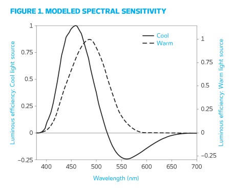
Figure 1. Circadian light is determined by these spectral weighting functions for cool and warm light sources.
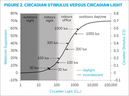
Figure 2. This graph derives from measurements of nocturnal melatonin suppression. Circadian stimulus levels at different photopic illuminance levels are also shown.
Circadian light is irradiance weighted by the spectral sensitivity of every retinal phototransduction mechanism that stimulates the biological clock, as measured by nocturnal melatonin suppression. Circadian stimulus is a transformation of circadian light into relative units, from zero (the threshold for circadian system activation) to 0.7 (response saturation), and is directly proportional to nocturnal melatonin suppression after one hour of light exposure (zero to 70 percent). Units and quantities for these metrics have been published in our 2010 Journal of Circadian Rhythms article “Circadian Light.” Predictions of melatonin suppression based on these metrics have been validated in the laboratory; these metrics have also been useful in describing light as it affects sleep and depression in several field studies, available on the Lighting Research Center (LRC) Light and Health website.
Lighting Research Center
Developed by the Lighting Research Center, the Daysimeter is worn at the eye level.
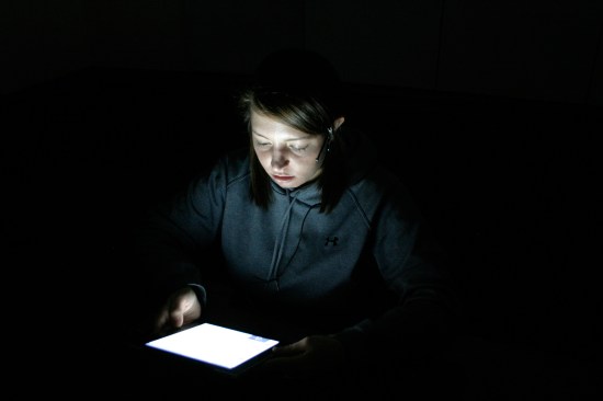
We measured the impact of light on acute nocturnal melatonin suppression in terms of circadian stimulus from iPads and computer screens, after a one-hour viewing time in two separate laboratory studies. We measured light incident at the cornea with the Daysimeter, a wearable device developed by LRC researchers in 2004 to measure the amount of circadian light a person receives during the study, and then compared the predicted suppression amount (corresponding to the circadian stimulus value) with the obtained result. In the iPad study, the predicted melatonin suppression of subjects was 3 percent and the measured melatonin suppression was also 3 percent. In the computer screen study, the predicted melatonin suppression was 23 percent and the measured value was 20 percent.
Lighting Research Center
Subjects in the iPad study were exposed to the display screen for one hour.
In terms of applications, six field studies we conducted showed that daytime light exposures of circadian stimulus greater than or equal to 0.3 are associated with better sleep, better mood, and lower depression. Perhaps of most interest, in three of those field studies, Alzheimer’s patients who received light exposures of circadian stimulus greater than or equal to 0.3 during daytime hours and less than 0.1 during the evening hours consistently and significantly increased their sleep duration, improved their sleep quality, and reduced their symptoms of depression and agitation.
Although the science underlying these advanced metrics may seem complex, the circadian light and circadian stimulus metrics can be applied with relative ease using our circadian stimulus calculator, available on the LRC Light and Health website, which enables a lighting professional to convert the photopic illuminance at the eye provided by any light source, at any light level, into the effectiveness of that light for stimulating the human circadian system.
Recommendations
Architectural lighting isn’t just for vision anymore. Clients are increasingly requesting and expecting lighting systems and applications that can support human health and well-being—and lighting professionals must be prepared to respond to those expectations. The first step must be measurement: If light as an enabler for health and well-being cannot be measured, it cannot be effectively delivered.
Although responses to circadian-effective light do vary from person to person, a lighting system that delivers a circadian stimulus greater than 0.3 during the day—particularly during the morning—and less than 0.1 in the evening is a great starting point. This guideline, of course, is overly simplistic. As such, we strongly encourage lighting professionals to seek opportunities that provide a deep understanding of the many ways light can affect health and well-being, and to become adept at addressing and designing lighting for special applications effectively. •
Select Resources
An introductory list of references and articles on the effect of light on the human circadian system.
Lighting Research Center Light and Health website.
“Light as a Circadian Stimulus for Architectural Lighting,” by Mark Rea and Mariana Figueiro, Lighting Research & Technology, 2016.
“Office Lighting and Personal Light Exposures in Two Seasons: Impact on Sleep and Mood,” by Mark Rea and Mariana Figueiro, Lighting Research & Technology, 2016.
“Light Level and Duration of Exposure Determine the Impact of Self-Luminous Tablets on Melatonin Suppression,” by Brittany Wood, et al., Applied Ergonomics, 2013.
“Comparisons of Three Practical Field Devices Used to Measure Personal Light Exposures and Activity Levels,” by Mariana Figueiro, et al., Lighting Research & Technology, 2013.