Our ongoing fascination with the brain has helped to spur the development of cognitive machines that replicate human thought processes. IBM researchers, for example, are working on neurosynaptic computing chips that emulate the structure of the human brain, while the new book Superintelligence: Paths, Dangers, Strategies (Oxford University Press, 2014), by Oxford University philosophy professor Nick Bostrom, outlines recent developments in artificial intelligence.
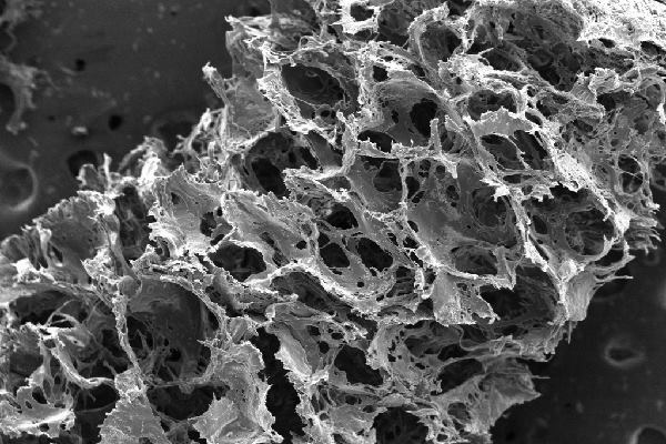
Researchers at the Tissue Engineering Resource Center (TERC) at Tufts University are also attempting to replicate brain functions. Instead of using software or hardware as their medium, they are working with bioengineered tissue. With funding from the National Institute of Biomedical Imaging and Bioengineering (NIBIB), the researchers fabricated 3D, living tissue similar in structure and function to that of rat brains. The composite tissue comprises two biomaterials: a gel made of collagen (white matter) that is surrounded by a silk protein–based spongy scaffold material (gray matter). The researchers cut the scaffold into a doughnut shape, injected it with rat neurons, and filled its center hole with gel, which permeated the scaffold. Within a few days, the neurons formed new networks within the scaffold and extended projections across the gel-filled center to the other side of the ring, effectively connecting the gel and scaffolding material.
“This work is an exceptional feat,” said TERC director Rosemarie Hunziker in a press release. “It combines a deep understanding of brain physiology with a large and growing suite of bioengineering tools to create an environment that is both necessary and sufficient to mimic brain function.”
The pseudo-brain tissue can stay alive for more than two months, allowing researchers to study the consequences of drug administration and traumatic brain injury more easily than with the actual brain tissue of living patients, which is difficult to evaluate before and after trauma in a controlled setting. “With the system we have, you can essentially track the tissue response to traumatic brain injury in real time,” said Tufts engineering professor David Kaplan. “Most importantly, you can also start to track repair and what happens over longer periods of time.”
The new surrogate material promises to increase scientists’ knowledge of the original brain. “Good models enable solid hypotheses that can be thoroughly tested,” Hunziker said. “The hope is that use of this model could lead to an acceleration of therapies for brain dysfunction as well as offer a better way to study normal brain physiology.”
Like other investigations into self-assembling materials, the Tufts research suggests provocative possibilities for architecture. Building systems may one day emulate those of living organisms, autonomously constructing and repairing themselves as needed. Perhaps future electrical networks will be made of bioengineered neurological tissue which, like the rat neurons in this experiment, will extend throughout architectural scaffolds, forming new connections on demand.
Blaine Brownell, AIA, is a regularly featured columnist whose stories appear on this website each week. His views and conclusions are not necessarily those of ARCHITECT magazine nor of the American Institute of Architects.